what is liposuction?


“Lipo” (from the Greek “lipos) means fat. And liposuction is a cosmetic surgical procedure by which excess fat is broken down and sucked out from under the skin. ​​Liposuction can be used to target fat under the chin, neck, cheeks, upper arms, breasts, abdomen, buttocks, hips, thighs, knees, calves, and ankle areas.

When performed by our expert plastic surgeons, liposuction can help reshape your contours – body and face. While it is certainly not a shortcut to drastic and instant weight loss, It eliminates those stubborn pockets of fat and restores youthful definition where it matters most: your midsection, thighs, flanks, and arms.


Widget not in any sidebars
Non-Thermal-Low-Level-Laser-as-an-aid-to-immunity-building-for-the-seasonal-nasties-seen-in-healthcare-and-clinical-practice.jpg

March 2, 2021

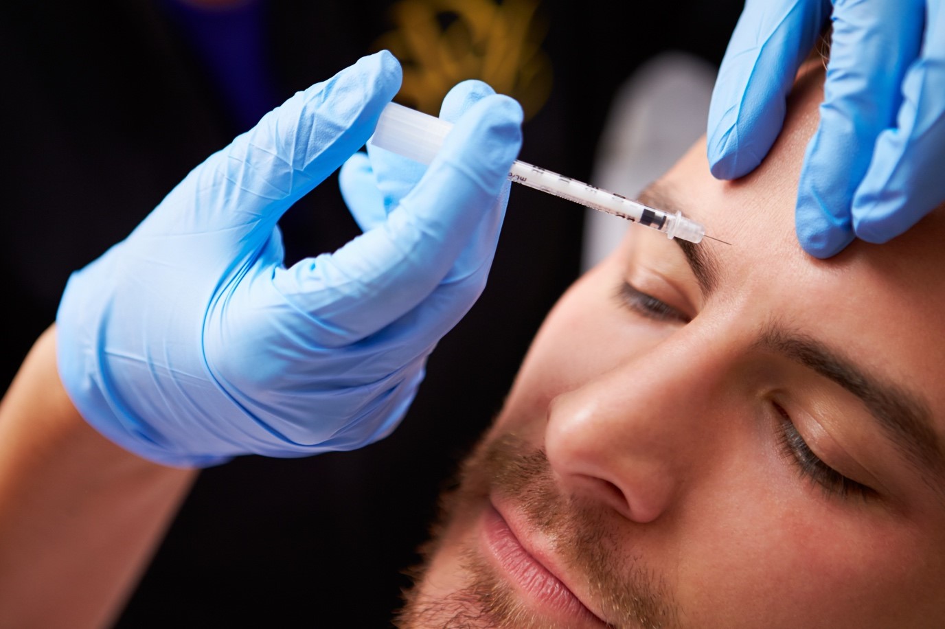
From autumn to spring we normally see colds, flu, aches and pains and a collection of seasonal nasties in clinical practice; no matter what discipline you practice in. if you have a non-thermal low-level laser, now is the time to put it so good use for you, your family, staff and clients. Have a read of this open literature review and see what conclusions you can draw. Given that this is 2020, this review leads with what’s topical, the evidence however is far reaching

This paper looks at the available research on using Non-thermal Low-Level laser as a therapy (NTLLLT), to improve and maintain a good immune response and is based on evidence, mindful of the current world situation in relation to Covid-19 and suggests that NTLLLT may be a viable intervention at this time for viral related seasonal nasties. This paper is not claiming the NTLLLT is a cure for Covid-19 or any disease, it’s merely based on what the evidence suggests.

Two strains of the new coronavirus that are spreading around the world, according to an analysis of 103 cases. But the World Health Organization insists that “there is no evidence that the virus has been changing”, viruses are intelligent and constant change and evolve.

Viruses are always mutating, especially RNA viruses like coronavirus SARS-CoV-2, and others. When a person is infected with the coronavirus, it replicates in their respiratory tract. Every time it does, around half a dozen genetic mutations occur, says Ian Jones at the University of Reading, UK.

When Xiaolu Tang et al at Peking University in Beijing studied the viral genome taken from 103 cases, they found common mutations at two locations on the genome. They identified two types of the virus based on differences in the genome at these two regions: 72 were considered to be the “L-type”(newer type), and 29 were classed “S-type” (older type).

A later analysis by Xiaolu Tang suggests that the L-type was derived from the older S-type. The first strain is likely to have emerged around the time the virus jumped from animals to humans, (anecdotally, there is a belief among some clinicians and members of the public, that this is not possible). The second type emerged soon after that species jump, according to Xiaolu et al. Both, we know, are involved in the current global outbreak. The fact that the L-type is more prevalent suggests that it is “more aggressive” than the S-type, the team say1.

Oxford Brookes University’s Ravinder Kanda, in the UK. Suggests that,  “The L-type might be more aggressive in transmitting itself, but we have no idea yet how these underlying genetic changes will relate to disease severity,”  Erik Volz at Imperial College London, in the same article says “I think it’s a fact that there are two strains” say “It’s normal for viruses to undergo evolution when they are transmitted to a new host.  The differences between the two identified strains are tiny.”

Coronaviruses are naturally hosted and evolutionarily shaped by bats and have been with us for a very long time. Indeed, it has been postulated that most of the coronaviruses in humans are derived from the bat reservoir. It is vital to know how many strains of the virus exist.

Coronaviruses were discovered in the mid 1960s by Tyrrell and Bynoe. The earliest discovered were an infectious bronchial virus in chickens and two in human pediatric patients who had what it was thought to be a common cold.  This was later named human coronavirus 229E and human coronavirus OC43. Other coronavirus type have been identified, these being, SARS-CoV in 2003, HCoV NL63 in 2004, HKU1 in 2005, MERS-CoV in 2012, and SARS-CoV-2 (formerly known as 2019-nCoV) in 2019. Most of these have involved serious respiratory tract infections.

Coronaviruses are large pleomorphic spherical particles with bulbous surface projections that look like a crown (corona). The diameter of the virus particles is around 120 nm. The envelope of the virus in electron micrographs appears as a distinct pair of electron dense shells.

The viral envelope consists of a lipid bilayer where the membrane, envelope and spike structural proteins are anchored. A subset of coronaviruses, specifically the members of Beta Coronavirus subgroup A, also have a shorter spike-like surface protein called hemagglutinin esterase (HE). Inside the envelope, there is the nucleocapsid, which is formed from multiple copies of the nucleocapsid protein, which are bound to the positive-sense single-stranded RNA genome in a continuous beads-on-a-string type structure or conformation. The lipid bilayer envelope, membrane proteins, and nucleocapsid protect the virus when it is outside the host cell.

Around the world, multiple groups are working on a vaccine for the virus. Any vaccine will need to target features that are found in both strains of the virus in order to be effective. Most promisingly, two drugs given together to treat HIV – called lopinavir and ritonavir – are already approved for human use, and in small trials they seemed to reduce disease severity and fatalities in people infected by the SARS or MERS coronaviruses (by reduction in the viral load). Doctors in Wuhan, the centre of the outbreak, have already started a randomised controlled trial of lopinavir and ritonavir. Covid-19 contains a strange HIV-like mutation that may make it more contagious and give it properties not found in other coronaviruses.

Based on what we know of HIV treatments and the links that have been drawn to it and the COVID-19 virus, it could be argued that the use of Non-thermal Low-Level Laser would have the same effect on coronavirus (by load reduction), as on HIV. Reactive species are frequently formed after viral infections. Antioxidant defences, including enzymatic and nonenzymatic components, protect against reactive species, but sometimes these defences are not completely adequate.

Oxygen radicals and nitric oxide (NO) are generated in excess in a diverse array of microbial infections. Emerging concepts in free radical biology are now shedding light on the pathogenesis of various diseases15, 16. Free‐radical induced pathogenicity in virus infections is of great importance, because evidence suggests that NO and oxygen radicals such as superoxide are key molecules in the pathogenesis of various infectious diseases. Although oxygen radicals and NO have an antimicrobial effect on bacteria and protozoa, they have opposing effects in virus infections such as influenza virus pneumonia and several other neurotropic virus infections.

An imbalance in the production of reactive species and the body’s inability to detoxify these reactive species is referred to as oxidative stress.

There is strong evidence which suggests that HIV-1 infected patients are under chronic oxidative stress, as are most viral infected patients. Thus, ROS has been suggested to be responsible for many aspects of HIV-1 pathogenesis such as increase viral replication, reduced immune cell proliferation, loss of immune function, and sensitivity to drug toxicity and chronic weight loss. Furthermore, excessive production of ROS can result in oxidation of proteins, peroxidation of lipids (seen in COVID-19), and eventually cell death. Non-thermal Low-Level Laser Therapy (NTLLLT), can improve the activity of antioxidant enzymes through a photochemical process that accelerates the elimination of ROS. This can be achieved at a molecular level by altering the conformation of antioxidant enzymes. A study conducted by Yang et al showed that LLLT (532 nm) can enhance the activity of anti-oxidant enzymes and also induce production of more ROS, with the amount produced dependent on the dose of the laser irradiation. Lugongolo et al in their paper on the treatment of HIV-1 suggest the use of 660 nm and also refer to a blue laser.

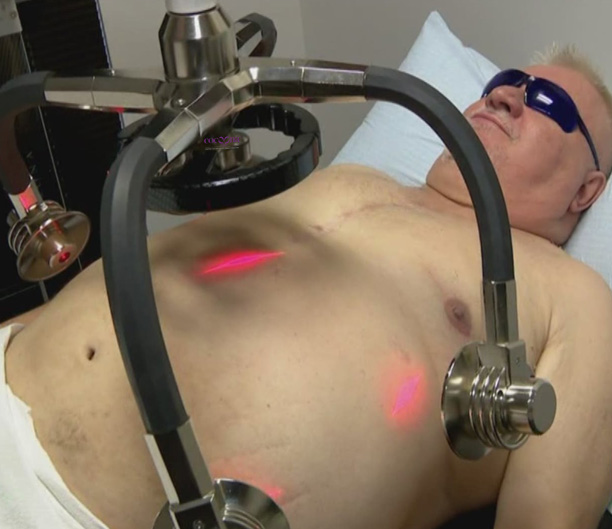
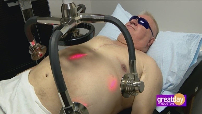
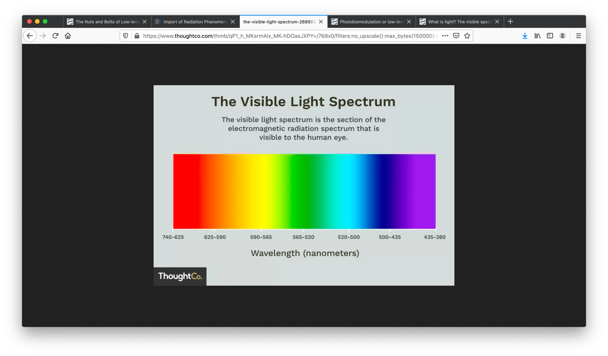
Non-thermal low-level lasers offer a collection of wavelengths, 400nm (blue violet), 530 nm (green), and 630 nm (red) and above. Lugongolo et al refer to similar frequencies.

Lugongolo et al, demonstrated the effects of laser irradiation in HIV-1 infected and uninfected TZM-bl cells. In addition, they showed that uninfected TZM-bl cells were stimulated by laser irradiation, while the effects of both HIV-1 infection and irradiation had detrimental effects on the cells. The TZM-bl cell line used in this study is a HeLa cell clone containing the CXCR4, CD4 and CCR5, which are host cell molecules the virus uses to gain entry into cells and making TZM-bl cell line permissive to HIV-1 infection. The TZM-bl cell line also contains a Tat-responsive firefly luciferase gene under the control of HIV-LTR, which gets expressed during HIV infection.

Herpes zoster, also known as shingles, produces a painful vesicular rash that results from the reactivation of the varicella zoster virus (VZV) treated with 632 nm responds in a similar way to HIV when irradiated using NTLLLT at 660 -880 nm. Both of these viruses respond to NTLLLT. 405 nm laser has also been investigated in the treatment of viral conditions.

An early study in 1991 by Skobelkin et al; performed preoperative NTLLLT on selected cancer patients undergoing palliative surgery. The levels of T-lymphocytes, T-helpers and Tsuppressors were assayed for the 7 days following the surgery, as were immunoglobulin levels, specifically IgA, IgM and IgG. The levels of blood-borne leukocytes, lymphocytes and monocytes all rose after laser therapy. Significantly increased levels of activated Tlymphocytes and helper T-cells were seen, with a significantly lower number of T-suppressors especially by the fifth post NLLLT day. Increased levels of IgA and IgG were seen by the second day, with a sharp reduction to almost normal levels by the fifth day. IgM levels rose slowly over the first four days, then rose sharply on the fifth day and maintained a high level during the period of the study. Skobelkin et al, proposed that these were all indications of a strong photoactivated immunological response, and boosting the competency of the immunocompetent systems of these long-term cancer patients. The high levels of IgG, especially cytotoxic for tumoural cells, has also been associated with a corresponding rise in killer T-cells. the antigen which would normally trigger these reactions was shown to be absent in all patients, thus the reaction was entirely photoactivated. Skobelkin et al, did not report any activation of tumoural remnants following LLLT, which has been of major concern to many researchers.

It could be concluded that utilising NTLLLT in in a proactive way, as a support to immunity, could be generally beneficial to the population, and in so doing give an immune boost to protect from the seasonal nasties. Consider it as a laser flu shot or an immune infusions. Our bodies have everything we need for a happy healthy life, all we sometime lack is the energy to be well, NTLLLT bring the energy to our cells that they need and can use to boot our immunity.


Reference:

  1. Xiaolu Tang, Changcheng Wu, Xiang Li, Yuhe Song, Xinmin Yao, Xinkai Wu, Yuange Duan, Hong Zhang, Yirong Wang, Zhaohui Qian, Jie Cui, Jian Lu, On the origin and continuing evolution of SARS-CoV-2, National Science Review, , nwaa036, https://doi.org/10.1093/nsr/nwaa036
  2. https://www.newscientist.com/term/coronavirus; 08-03-2020 verified 09-03-2020
  3. Prof Ian Jones, 2020; University of Reading, BBC interview 04-03-2020
  4. New Scientist, 22nd January 2020. Wuhan coronavirus may have been transmitted to people from snakes. Article by Jessica Hamzelou
  5. New Scientist, 5th March 2020. Coronavirus: Are there two strains and is one more deadly? Article by Jessica Hamzelou Cui J, Li F, Shi Z-L.
  6. Origin and evolution of pathogenic coronaviruses. Nature Reviews Microbiology. 2019;17(3):181-92. doi:1038/s41579-018-0118-9.
  1. Li X, Song Y, Wong G, Cui J. Bat origin of a new human coronavirus: there and back again. Science China Life Sciences. 2020. doi: 10.1007/s11427-020-1645-7.
  1. Li W, Shi Z, Yu M, Ren W, Smith C, Epstein JH, et al. Bats are natural reservoirs of SARS-like coronaviruses. Science. 2005;310(5748):676-9. Epub 2005/10/01. doi: 10.1126/science.1118391. PubMed PMID: 16195424.Downloaded from https://academic.oup.com/nsr/advance-article-abstract/doi/10.1093/nsr/nwaa036/5775463 by guest on 08 March 202
  2. Dominguez SR, O’Shea TJ, Oko LM, Holmes KV. Detection of group 1 coronaviruses in bats in North America. Emerg Infect Dis. 2007;13(9):1295-300. Epub 2008/02/07. doi: 10.3201/eid1309.070491. PubMed PMID: 18252098; PubMed Central PMCID: PMCPMC2857301
  3. thelancet.com/journals/lancet/article/PIIS0140-6736(20)30183-5/fulltext 24-01-2020; verified 08-03-2020
  4. ccn.com/hiv-ebola-like-mutations-suggest-coronavirus-leaked-from-a-lab/
  5. Lugongolo, Masixole & Manoto, Sello & Ombinda Lemboumba, Saturnin & Maaza, Malik & Mthunzi-Kufa, Patience. (2017). The effects of low level laser therapy on both HIV-1 infected and uninfected TZM-bl cells. Journal of Biophotonics. 10. 1002/jbio.201600217.
  1. Camini, F.C., da Silva Caetano, C.C., Almeida, L.T. et al. Implications of oxidative stress on viral pathogenesis. Arch Virol 162, 907–917 (2017). https://doi.org/10.1007/s00705-016-3187-y
  2. Yashoda Madaiah Hosakote and Kempaiah Rayavara, Respiratory Syncytial Virus-Induced Oxidative Stress in Lung Pathogenesis, Oxidative Stress in Lung Diseases, 10.1007/978-981-32-9366-3_13, (297-330), (2019).
  3. ranger DL, Hibbs JB Jr, Perfect JR,et al. Specificamino acid (L-arginine) requirement for microbio-static activity of murine macrophages. JClin Invest1988;81: 1129–1136
  4. Badwey JA, Karnovsky ML. Active oxygen species and the functions of phagocytic leukocytes.AnnuRev Biochem1980;49:695–726.
  1. Moncada S, Higgs A. The L-arginine–nitric oxide pathway.N Engl JMed1993;329: 2002–2012.
  2. Akaike T, Yoshida M, Miyamoto Y,et al. Antag-onistic action of imidazolineoxylN-oxides againstendothelium-derived relaxing factor/eNO througha radical reaction.Biochemistry1993;32
  3. Akaike T, Maeda H. Nitric oxide and virus infection. Immunology. 2000;101(3):300–308. doi:10.1046/j.13652567.2000.00142.x
  4. L. Ngondi, J. Oben, L. H. Etame, D. M. Forkah, and D. Mbanya, AIDS Research Therapy 3, 19 (2006).
  5. M. V. Reiche and A. N. C. Simão, in: Dr. Nancy Dumais (ed.) Updates on Biology, Immunology, Epidemiology and Treatment Strategies (2011), pp. 24–68.
  6. F. Yang, J. G. Yang, G. H. Gao, Z. R. Hu, H. X. Chen, and H. W. Qian, Acta Laser Biology Sinica 11, 388–394 (2002).
  7. Lugongolo, M.Y., Manoto, S.N., Ombinda-Lemboumba, S., Maaza, M., & Mthunzi-Kufa, P. (2017). The effects of low level laser therapy on both HIV-1 infected and uninfected TZM-bl cells. Journal of biophotonics, 10 10, 1335-1344 .
  8. J. Platt, K. Wehrly, S. E. Kuhmann, B. Chesebro, and B. D. Kabat, J. Virol. 72, 2855–2864 (1998).
  9. R. Mascola, P. D’Souza, P. Gilbert, B. Hahn, N. L. Haigwood, L. Morris, J. C. Petropoulos, V. R. Polonis, M. Sarzotti-Kelsoe, and D. C. Montefiori, J. Virol. 79, 10103–10107 (2005).
  10. Edelsburg JS, Lord C, Oster G. Systematic review and meta-analysis of efficacy, safety and tolerability data from randomized controlled trials of drugs used to treat postherpetic neuralgia. Ann Pharmacother. 2011;45(12):1483-1490.
  11. Dubinksy RM, Kabani H, El-Chami Z. Practice parameter: treatment of postherpetic neuralgia: An evidence based report of the quality standards subcommittee of the American Academy of Neurology. Neurology. 2004;63(6):959-965.
  12. Simunovic, z. (2012). Herpes virus infection low level laser therapy (lllt)- photobiostimulation applied as mono therapy in treatment of human pathogen herpes virus.
  13. Tomb RM, Maclean M, Coia JE, et al. New Proof-of-Concept in Viral Inactivation: Virucidal Efficacy of 405 nm Light Against Feline Calicivirus as a Model for Norovirus Decontamination. Food Environ Virol. 2017;9(2):159–167. doi:10.1007/s12560016-9275-z
  1. Kahn, Jeffrey; McIntosh, Kenneth (November 2005), “History and recent advances in coronavirus discovery”, Pediatric Infectious Disease Journal, 24 (11): s223–s227, doi:10.1097/01.inf.0000188166.17324.60, archived from the original on 5 February 2020
  2. Geller C, Varbanov M, Duval RE (November 2012). “Human coronaviruses: insights into environmental resistance and its influence on the development of new antiseptic strategies”. Viruses. 4 (11): 3044–68. doi:10.3390/v4113044. PMC 3509683. PMID 23202515.
  3. Goldsmith CS, Tatti KM, Ksiazek TG, Rollin PE, Comer JA, Lee WW, et al. (February 2004). “Ultrastructural characterization of SARS coronavirus”. Emerging Infectious Diseases. 10 (2): 320–6. doi:10.3201/eid1002.030913. PMC 3322934. PMID 15030705.
  4. Fehr AR, Perlman S (2015). Maier HJ, Bickerton E, Britton P (eds.). “An Overview of Their Replication and Pathogenesis; Section 2 Genomic Organization”. Methods in Molecular Biology. Springer. 1282: 1–23. doi:10.1007/978-1-4939-2438-7_1. ISBN 978-1-4939-2438-7. PMC 4369385. PMID 25720466. See section: Virion Structure.
  5. Neuman BW, Adair BD, Yoshioka C, Quispe JD, Orca G, Kuhn P, et al. (August 2006). “Supramolecular architecture of severe acute respiratory syndrome coronavirus revealed by electron cryomicroscopy”. Journal of Virology. 80 (16): 7918–28. doi:10.1128/JVI.00645-06. PMC 1563832. PMID 16873249. Particle diameters ranged from 50 to 150 nm, excluding the spikes, with mean particle diameters of 82 to 94 nm.
  6. Lai MM, Cavanagh D (1997). “The molecular biology of coronaviruses”. Advances in Virus Research. 48: 1–100.

doi:10.1016/S0065-3527(08)60286-9. ISBN 9780120398485. PMID 9233431.

  1. de Groot RJ, Baker SC, Baric R, Enjuanes L, Gorbalenya AE, Holmes KV, Perlman S, Poon L, Rottier PJ, Talbot PJ, Woo PC,

Ziebuhr J (2011). “Family Coronaviridae”. In King AM, Lefkowitz E, Adams MJ, Carstens EB, International Committee on Taxonomy of Viruses, International Union of Microbiological Societies. Virology Division (eds.). Ninth Report of the International Committee on Taxonomy of Viruses. Oxford: Elsevier. pp. 806–28. ISBN 978-0-12-384684-6.

  1. Chang CK, Hou MH, Chang CF, Hsiao CD, Huang TH (March 2014). “The SARS coronavirus nucleocapsid protein–forms and functions”. Antiviral Research. 103: 39–50. doi:10.1016/j.antiviral.2013.12.009. PMID 24418573.
  2. Neuman BW, Kiss G, Kunding AH, Bhella D, Baksh MF, Connelly S, et al. (April 2011). “A structural analysis of M protein in coronavirus assembly and morphology”. Journal of Structural Biology. 174 (1): 11–22. doi:10.1016/j.jsb.2010.11.021. PMC 4486061. PMID 21130884.
  3. Tyrrell DA, Bynoe ML. Cultivation of viruses from a high proportion of patients with colds. Lancet. 1966;1:76–77.
  4. Skobelkin, O.K., Michailov, V.A., & Zakharov, S.D. (1991). Preoperative activation of the immune system by low reactive level laser therapy (lllt) in oncologic patients : a preliminary report.

Dr. Sanjay Parashar

Non-Thermal-635nm-Low-Level-Laser-Therapy-in-Pre-Diabetes-and-Obesity-Management.jpg

March 2, 2021

Background

Pre-Diabetes and pre-obesity often go hand in hand. Diabetes mellitus (DM) is a chronic condition that can alter our carbohydrate, protein, and fat metabolism. It is caused by the absence of insulin secretion due to either the progressive or marked inability of the β-Langerhans islet cells of the pancreas to produce insulin, or due to defects in insulin uptake in the peripheral tissue. DM is broadly classified under two categories, which include type 1 and type 2 diabetes.

Body mass index has a strong relationship to diabetes and insulin resistance. In obese individuals, the amount of non-esterified fatty acids (NEFA), glycerol, hormones, cytokines, proinflammatory markers, and other substances that are involved in the development of insulin resistance, is increased.

The pathogenesis in the development of diabetes is due to the β-islet cells of the pancreas becoming impaired, causing a lack of control of blood glucose. The development of diabetes becomes more inevitable if the failure of β-islet cells of the pancreas is accompanied by insulin resistance. Weight gain and body mass are central to the formation and rising incidence of type 1 and type 2 diabetes.

In conclusion, new approaches in managing and preventing diabetes in obese individuals must be studied and investigated based on the facts. True Non-Thermal Low-Level Laser (TNTLLL)may form part of the solution.

The association between type 1 diabetes and weight gain was first investigated by Baum et al in 1975. The Baum et al study suggested that there was an association related to overfeeding or to hormonal dysregulation.

Overweight and obesity are defined by an excess accumulation of adipose tissue which impairs both physical and psychosocial health and well-being. Obesity is considered a health disaster in both developed and developing countries.

The increased prevalence of obesity in the current climate has drawn attention to the worldwide significance of this problem. In the US, approximately two-thirds of the adult population is overweight or obese and similar trends are being noticed worldwide. Obesity is linked to many medical, psychological, and social conditions, the most devastating of which may be type 2 diabetes. At the start of this century, 171 million people were estimated to have type 2 diabetes, and this figure is expected to increase to 360 million by 2030. The figures from the World Health Organisation show that around 422 million people worldwide have diabetes, with the majority living in low-and middle-income countries, and 1.6 million deaths are directly attributed to diabetes each year. Both the number of cases and the prevalence of diabetes have been steadily increasing over the past few decades. A comparison of the two sets of statistics shows just what a combined problem these two conditions represent. The Pharmacoeconomics, not to mention the associated costs on health services is staggering. In 2016 the estimated burden of diabetes on healthcare infrastructure was 825 Billion USD. There are no published figures for 2020, but it is estimated to be more than a trillion USD.

The Study

The study enrolled 140 men and women between the ages of 18 to 70 with a body mass index (BMI) of 27 to 45. This was a 24 month study including follow up. Participating subjects were randomised in a double-blind fashion to receive non-thermal low-level laser. The study was set up as below.

Non-esterified fatty acids (NEFA) are molecules released from triglycerides by the action of the enzyme lipase and are transported in the blood bound to albumin. They contribute only a small proportion of the body’s fat; however, provide a large part of the body’s energy. Measurement of NEFA is important in diabetes where insulin deficiency results in the metabolism of fat. Levels are also frequently increased in obese patients.

Subject sample

Active Subjects Placebo Subjects Total Subjects
70 70 140

Subject demographics

Gender

Group Male Female Total
Active 35 (50%) 35 (50%) 70 (50%)
Placebo 37 (53%) 33 (47%) 70 (50%)

Age

Group Active Placebo Total
Mean 50.13 49.24 49.69
SD 9.78 9.78 9.76
Range 35 – 65 33 – 70 33 – 70

A t-test for independent samples reported a difference of 0.89 years between randomized subjects to the active and placebo treatment groups to be not statistically significant (t=+0.54, p=0.59; p>0.05).

All subjects involved in this study were recommended and referred by their medical team and had to be pre-diabetic and pre-obese or obese and not taking medication for these conditions.

Treatment device.

The low-level laser energy device is a non-invasive dermatological aesthetic treatment cleared by the FDA for use as a non-invasive aesthetic dermatological treatment for reducing the circumference of hips, waist, and thighs and is manufactured by Erchonia Corporation, USA. The LLLT device consists of four independent diodes that are positioned 120 degrees apart and tilted at a 30-degree angle. A fifth diode is positioned at the centreline.

The 17mW of red 635nm of laser light emitted from each diode is collected and processed through a proprietary lens that redirects the beam with a line refractor. The refracted light from each diode is bent into a random spiralling circle pattern that is independent of the other diodes. These overlapping patterns ensure total coverage of the target treatment area. The total amount of energy delivered to the skin during each treatment as stated in the FDA clearance was 3.94J/cm2. Evidence however suggests that this is not relevant as the laser delivers photonic energy via electromagnetic energy transfer.

Procedure.

Each subject was randomised to receive 20 active or sham treatments 2 per week, equally spaced apart with the low-level energy laser device over a 10 week period. Both the active and sham devices have the same physical appearance and emit light when activated that is indistinguishable to both the subject and the administration investigator. All subjects had moderated exercise sessions after each treatment, and another on a day of their choice. Nutritional plans were also moderated for compliance.

Study assessments.

The circumference of each participant was measured using a flexible tape measure pre- and post- each application at 3 points; the base of the circumference at sternum, the circumference at the umbilicus, and again at the trochanter.  For accuracy at remeasuring post treatment, a skin marker was used under the tape at several points so the measurements were duplicated. These points were recorded at baseline to ensure that subsequent measurements were obtained at the same location. All measurements were performed by a member of the investigative team not involved in performing the actual treatments. All subjects were photographed from front, sides and back with hands on head.

The primary outcome measure was the number of subjects accumulated BMI and the total decrease achieved. Individually the three combined measurement points after each session were recorded. Blood glucose was also measured each week along with weight and other

variables such as sleep quality and energy levels. Secondary outcomes assessed at the completion of the study included changes in BMI, associated diabetes risk, obesity levels and several subjective ratings, which measured subject attitudes about overall satisfaction with their results and improvements in the appearance.

All study assessments were performed at baseline, at the completion of treatment, and two weeks post-treatment. Following the baseline physical examination, a blinded investigator noted any changes in existing skin condition including scars, cellulite, stretch marks, discoloration, stria, dimpling, skin quality and elasticity following treatment. Details about food and drink consumption, physical activity, and adverse events for each subject were recorded daily. Further follow ups were recorded every 12 weeks primarily for the first 18 months from inception, then extended over the 5 year period. The study is now concluded.

Ethics.

The protocol used in this study adhered to the Good Clinical Practice guidelines and was approved by the local ethics committee in April 2015. The study was overseen by colleagues in the diabetes assessment unit at the local university hospital.   Informed written consent was obtained from each subject prior to participation in any study-related activities.

Laser Diode Placement

The diodes are placed over the lateral flanks and around the umbilical area. The treatment was performed for 20 minutes and is repeated for a further 20 minutes, with the participant repositioned. A more comfortable method for a heavier subject is for them to lay on their side and the diodes positioned accordingly to cover half of the midsection, the subject then repositions to the other side.

Rational

To find an alternative to the current interventions for diabetes, obesity and associated conditions, to reduce the overall economic burden, and to improve quality outcomes and quality of life for sufferers.

Baseline variables

BMI

Group Active Placebo Total
Mean 5.09 5.62 5.35
SD 1.65 1.80 1.74
Range 3.1 – 8.8 3.1 – 9.1 3.1 – 9.1

A t-test of independent samples found the 0.53 difference in baseline Total Cholesterol between subjects randomized to the active and placebo treatment groups to be not statistically significant (t=-1.82, p=0.071; p>0.05).

Throughout the study all participants had their blood pressure, cholesterol and blood sugars monitored. Changes in behaviour were also considered as was sleep patterns, changes in energy levels and general skin appearance. A glycated haemoglobin test (HbA1c) was taken every 12 weeks.

Total Cholesterol

Group Active Placebo Total
Mean 5.09 5.62 5.35
SD 1.65 1.80 1.74
Range 3.1 – 8.8 3.1 – 9.1 3.1 – 9.1

A t-test for independent samples found the 0.53 difference in baseline Total Cholesterol between subjects randomized to the active and placebo treatment groups to be not statistically significant (t=-1.82, p=0.071; p>0.05).

HbA1c

Group Active Placebo Total
Mean 6.46 6.46 6.86
SD 0.18 0.16 4.70
Range 5.52 – 6.66 5.2 – 6.61 5.2 – 6.66

There is no difference (0 points) in baseline HbA1c between subjects randomized to the active and placebo treatment groups.

Comparisons across evaluations

The following evaluations of study variables were made:

  1. Baseline (pre-treatment)
  2. End of Treatment
  3. Follow-up

The following analyses evaluate the change in evaluations for each study measure across the three evaluations, as applicable.

BMI

BMI Baseline Treatment End Follow-Up
Active               Mean 33.41 30.54 25.86
                             SD 2.67 2.35 1.39
Placebo            Mean 33.57 31.86 36.40
                             SD 3.16 3.12 1.86

As this is a simple inhouse study, the information shared would benefit from more testing within a full RTC. The lead researcher and his team maintained strict protocols throughout this study (see appendix 1).

A one-way ANOVAs for 3 correlated samples was conducted to evaluate change in BMI across the study duration within each of the active and placebo treatment groups, with results as follows:

Active Group

There is a statistically significant difference in BMI ratings across the study evaluation duration for active group subjects (F=563.65, p<0.001). Subsequent Tukey Analysis revealed the statistically significant differences to have occurred between the following evaluations, at p<0.01:

  • Baseline to Treatment End
  • Baseline to Follow-Up
  • Treatment End to Follow-Up

Placebo Group

There is a statistically significant difference in BMI ratings across study evaluation duration for placebo group subjects (F=93.71, p<0.001). Subsequent Tukey Analysis revealed the statistically significant differences to have occurred between the following evaluations, at p<0.01:

  • Baseline to Treatment End
  • Baseline to Follow-Up
  • Treatment End to Follow-Up

In summary, both active and placebo subject groups evidenced a statistically significant decrease in BMI from pre-treatment to treatment end evaluation. However, while both groups likewise evidenced statistically significant changes in BMI from treatment end to follow-up evaluation, this change was in the direction of a statistically significant mean decrease for active group subjects and a statistically significant mean increase for placebo group subjects. By follow-up evaluation, active group subjects evidenced a mean decrease in BMI of 7.55. In contrast, placebo group subjects evidenced a mean increase in BMI of 2.83 across the same evaluation period.

Furthermore, while a t-test for independent samples found a 0.16 difference in baseline (pre-treatment) BMI between active and placebo subject groups to be not statistically significant (p>0.05), the 10.54 difference in BMI between active and placebo groups at follow-up evaluation was seen as statistically significant (p<0.0001).

HbA1c

HbA1c Baseline Treatment End Follow-Up
Active               Mean 6.46 5.66 4.68
                             SD 0.18 0.20 0.40
Placebo            Mean 6.46 6.10 9.56
                             SD 0.16 0.26 1.67

A one-way ANOVA for 3 correlated samples was conducted to evaluate change in HbA1c across study duration within each of the active and placebo treatment groups, with results as follows:

Active Group: There is a statistically significant difference in HbA1c ratings across study evaluation duration for active group subjects (F=797.77, p<0.0001). Subsequent Tukey Analysis revealed the statistically significant differences to have occurred between the following evaluations, at p<0.01:

  • Baseline to Treatment End
  • Baseline to Follow-Up
  • Treatment End to Follow-Up

Placebo Group: There is a statistically significant difference in HbA1c ratings across study evaluation duration for placebo group subjects (F=274.79, p<0.0001). Subsequent Tukey Analysis revealed the statistically significant differences to have occurred between the following evaluations, at p<0.01:

  • Baseline to Follow-Up
  • Treatment End to Follow-Up

In summary, similarly to the BMI findings, both active and placebo subject groups evidenced a decrease in HbA1c from pre-treatment to treatment end evaluation, although the decrease was only statistically significant for the active treatment group at p<0.01. However, while both groups evidenced statistically significant changes in HbA1c from treatment end to follow-up evaluation, this change was a statistically significant decrease for active group subjects and a statistically significant increase for placebo group subjects. By follow-up evaluation, active group subjects evidenced a statistically significant mean decrease

in HbA1c of 1.78 relative to baseline. In contrast, placebo group subjects evidenced a statistically significant mean increase in HbA1c of 3.10 across the same evaluation period.

Furthermore, while there was no difference in baseline (pre-treatment) HbA1c between active and placebo subject groups (both 6.46), the 4.88 difference in mean HbA1c between active and placebo groups at follow-up evaluation was statistically significant in favor of a significantly lower HbA1c for active group subjects relative to placebo group subjects (p<0.0001).

Total Cholesterol

Total Cholesterol Baseline Follow-Up
Active               Mean 5.09 3.53
                             SD 1.65 0.50
Placebo            Mean 5.62 5.76
                             SD 1.80 1.49

T-tests for correlated samples were conducted to evaluate change in total cholesterol across the study duration for each of the active and placebo treatment groups, with results as follows:

Active Group

T=10.11, p<0.0001. There is a statistically significant 1.56-point decrease in total cholesterol ratings from pre-treatment to treatment follow-up evaluation for active group subjects.

Placebo Group

There is no change in total cholesterol ratings from pre-treatment to follow-up evaluation for placebo group subjects (t=-1.32, p=0.19; p>0.05), with the 0.14-point increase being not statistically significant.

Furthermore, while there was no statistically significant difference in baseline (pre-treatment) total cholesterol between active and placebo subject groups (p>0.05), the 2.23-point difference in mean total cholesterol between active and placebo groups at follow-up evaluation was statistically significant (t=-11.89, p<0.0001).

Blood pressure

Blood pressure was recorded at baseline (pre-treatment), treatment end, and follow-up.

At baseline, blood pressure readings for all subjects in both the active and placebo groups were within the Hypertension Group 1 or Group 2 category.

Across treatment end and follow-up evaluations, all 70 subjects in the active treatment group demonstrated a progressive improvement (lowering) in blood pressure readings, and 12 active group subjects had lowered their blood pressure reading to within the pre-hypertension range by follow-up evaluation.

In contrast, there were no notable improvements in blood pressure readings for any of the 70 placebo group subjects from pre-treatment to follow-up evaluation, and no placebo group subject recorded a blood pressure rating below the Hypertension Stage 1 category at follow-up evaluation.

Sleep quality: P, A, I, G, E.

The P, A, I, G, E sleep quality scale used in this study is provided below.

P = You take more than 30 minutes to fall asleep after you get into bed.

You regularly wake up more than once per night.

You lie awake for more than 20 minutes when you wake up in the middle of the

night.

Feel tired quickly upon wakening.

A = You take less than 30 minutes to fall asleep after you get into bed.

You wake no more than once per night.

Your sleep is restless and your bed is tossed when you awaken.

Feel tired midday.

I = You take less than 20 minutes to fall asleep after you get into bed.

You wake no more than once per night.

You return to sleep quickly but sleep light.

Feel tired in the afternoon.

G = You fall asleep quickly after you get into bed.

You wake occasionally in the night.

You return to sleep quickly if you do awaken.

You seldom feel tired in the day.

E = You fall asleep quickly after you get into bed.

You do not wake in the night.

You awaken naturally and feel refreshed.

You never feel tired in the day.

Therefore, progression from ‘P’ through ‘E’ represents improvement in sleep quality.

Below is a summary of the percentage of subjects in each of the active and placebo groups, respectively, who rated their sleep quality on this scale as ‘P’, ‘A’, ‘I’, ‘G, or ‘E’ at each of the three evaluations.

Active Group Baseline Treatment End Follow-up
P 30 (43%) 1 (1%)
A 29 (41%) 12 (17%)
I 37 (53%) 9 (13%)
G 11 (16%) 12 (17%) 13 (19%)
E 9 (13%) 47 (67%)

At basepoint evaluation, eighty-four per cent (84%) of active group subjects rated their sleep quality as ‘P’ or ‘A’ – the two poorest sleep quality categories. By Follow-Up evaluation, only one active group subject reported poor sleep quality, and each of the remaining active group subjects indicated improved sleep quality, with 86% of active group subjects reporting their sleep quality as ‘G’ or ‘E’ – the two best sleep quality categories.

Placebo Group Baseline Treatment End Follow-up
P 39 (56%) 20 (29%) 21 (31%)
A 24 (34%) 37 (53%) 19 (28%)
I 11 (16%) 13 (19%)
G 7 (10%) 2 (2%) 15 (22%)
E

Similarly to active group subjects, 90% of placebo group subjects rated their sleep quality as ‘P’ or ‘A’ – the two poorest sleep quality categories – at baseline evaluation. However, by Follow-Up evaluation, 59% of placebo group subjects continued to record a ‘P’ or ‘A’ rating compared with the single active group subject, and only 22% of placebo group subjects reported their sleep quality as ‘G’ or ‘E’ – the two best sleep quality categories at follow-up evaluation compared with 86% of active group subject – almost four times fewer.

At the end of the 10 week active phase, all the subjects in the active protocol had undergone 4 weeks of further monitoring in relation to their blood glucose levels, and 69 of them no longer presented as a diabetes risk. The control group however all remained on the diabetes index as being at risk at week 10 +4.  All subjects were encouraged to follow their established exercise routine and to foster their nutritional habits. Follow-ups were taken for 24 months.

In the control group 69 went on to develop diabetes and one died because of complications related to diabetes. Over time their weight increased. The female members of the group maintained reduced weight and cholesterol levels. All of the subjects continued with exercises and a reasonable diet.

Discussion

There is a growing body of research showing the benefits of NTLLL in the management of the human fat cell, pain management and neurological disorders but as yet there is no substantive research into the benefits of this technology in the management of obesity and diabetes. Weight contributed to rheumatoid arthritis, heart disease, osteoarthritis, is now a contributory factor in problems associated with the coronavirus. There are well recorded positive side effects associated with the use of NTLLL[i], and these effects need to be exploited. This is a simple inhouse study exploring the use of NTLLL at 635nm in pre-diabetes/pre-obese patients.  Can we do anything to reduce the potential development of diabetes?

Conclusion

Diabetes is potentially a life limiting condition and fraught with many comorbidities.  Patients may go on to develop issues with blood pressure, kidneys, neuropathies, and amputations. The list is practically endless. This study, though inhouse, has followed best practice in terms of RTC, and though it is not perfect, the evidence presented speaks for itself. NTLLL may have a place in the armament of treatments used to prevent and control what is one of the world’s biggest health issues. 65 out of 70 participants in the study who were pre-diabetic and pre-obese, and in a 10 week period, turned their lives around. Over a 5 year period they continued to improve and maintained the good habits they had formed on the study.  They were no longer a diabetes risk and were not obese, and they all had good cholesterol levels and no blood pressure issues. They are no longer a burden on the healthcare systems. Although further studies are required, NTLLL could have long lasting benefits for diabetes, obesity, and health generally.


Reference:

  1. Scheen AJ. Pathophysiology of type 2 diabetes. Acta Clin Belg. 2003;58(6):335–341.
  2. van Belle TL, Coppieters KT, von Herrath MG. Type 1 diabetes: etiology, immunology, and therapeutic strategies. Physiol Rev. 2011;91(1):79–118.
  3. Al-Goblan, A. S., Al-Alfi, M. A., & Khan, M. Z. (2014). Mechanism linking diabetes mellitus and obesity. Diabetes, metabolic syndrome and obesity : targets and therapy, 7, 587–591. https://doi.org/10.2147/DMSO.S67400
  4. Baum JD, Ounsted M, Smith MA. Letter: Weight gain in infancy and subsequent development of diabetes mellitus in childhood. Lancet. 1975;2(7940):866.
  5. Naser KA, Gruber A, Thomson GA. The emerging pandemic of obesity and diabetes: are we doing enough to prevent a disaster? Int J Clin Pract. 2006;60(9):1093–1097.
  6. Gallagher D, Heymsfield SB, Heo M, Jebb SA, Murgatroyd PR, Sakamoto Y. Healthy percentage body fat ranges: an approach for developing guidelines based on body mass index. Am J Clin Nutr. 2000;72(3):694–701.
  7. Arora S. Insulin Resistance. Rijeka, Croatia: InTech Europe; 2012. [Accessed September 26, 2014]. Molecular basis of insulin resistance and its relation to metabolic syndrome.
  8. Tsai AG, Williamson DF, Glick HA. Direct medical cost of overweight and obesity in the USA: a quantitative systematic review. Obes Rev. 2011;12(1):50–61.
  9. McKeigue PM, Shah B, Marmot MG. Relation of central obesity and insulin resistance with high diabetes prevalence and cardiovascular risk in South Asians. Lancet. 1991;337(8738):382–386.
  10. https://www.who.int/health-topics/diabetes#tab=tab_1
  11. https://www.hsph.harvard.edu/news/press-releases/diabetes-cost-825-billion-a-year/
  12. Farivar, S., Malekshahabi, T., & Shiari, R. (2014). Biological effects of low level laser therapy. Journal of lasers in medical sciences, 5(2), 58–62.

Dr. Sanjay Parashar

Understanding-Non-Thermal-Low-Level-Lasers-Its-not-all-about-penetration.jpg

March 2, 2021

Put simply, Non-Thermal Low-Level Laser (NTLLL) delivers energy to the mitochondria of cells. This energy is delivered as photons or light particles, and this process is referred to as electromagnetic energy transfer. The aim of this article is to provide a brief synopsis to help the reader understand electromagnetic waves (EMW) and NTLLL.

Electromagnetic energy is a type of energy that is able to travel at the speed of light, it is characterised as having both electric and magnetic fields.

From humble beginnings more than 100 years ago, EMR has become an important component of modern medicine. There is an urgent need for education and better understanding with respect to its principles and applications.

The application of this energy is not new, Endre Mester at the Semmelweis Medical University in Hungary understood its effect.  In 1998, Wilden showed the importance of low-level laser in the delivery of energy.  Wilden went on to say, “Depending on its wavelength, electromagnetic radiation in the form of light can stimulate macromolecules and can initiate conformation changes in proteins or can transfer energy to electrons. Low level laser from the red and the near infrared region corresponds well with the characteristic energy and absorption levels of the relevant components of the respiratory chain”. What he was talking about is Non-Thermal Electromagnetic Transfer via Light. NTLLL is a collimated, monochromatic, unidirectional beam of light from the visual light spectrum. The visible spectrum is the portion of the electromagnetic spectrum that is visible to the human eye.

When you take visual light from the electromagnetic radiation spectrum, depth of penetration in terms of NTLLL becomes totally irrelevant. It is not about penetration, it’s about the wave. The concept of a wave is very familiar to all of us.  We have all seen waves travel across the beach, some are short and fast, while others are fast and slower, and some are just ripples. Electromagnetic radiation/Visual light, is very much like that.

Electromagnetic radiation is part of our everyday lives.  These waves are penetrating our bodies 24 hours a day, 7 days a week, and they never stop. Life could not function without them. Electromagnet waves have many characteristics, and their two most fundamental are wavelength and frequency. Figure 1.1 shows a sinusoidal electromagnetic wave in general. The direction axis of the wave is marked by z this is sometime called the  k-vector. EM waves have two oscillating parts, one electric the

other magnetic (x and  y in the diagram). The magnetic field is at right angles to the electric field and vice versa.

As can be seen from the above diagram, the wavelength is the distance (nanometres nm) between two adjacent peaks or troughs. This distance is measured along the axis z.  How many times per second the wave oscillates is the frequency. EM waves pass through the body without causing any damage.

When we relate the above information to NTLLL, and the fact that they operate from the visual light part of the electromagnetic spectrum; they use minimal power to generating a stable laser beam, and this beam consists of an electromagnetic wave that passes through the body.  Depth of penetration is irrelevant.

There are hundreds of so called lasers out there, and some are lasers, whilst some are not. A True laser is a device that generates an intense beam of coherent monochromatic light (or electromagnetic radiation) by stimulated emission of radiation (photons) from excited atoms or molecules.  Any device claiming to be a laser must exhibit these qualities.

Our bodies rely on photons to maintain good health. Energy from photons or light particles can be absorbed or released by electrons. When an electron absorbs a photon, the energy can free the electron to move around, or the electron can release the energy as another photo. Biophotons are light particles that are generated within the body and are constantly radiated from the body surface. These spontaneous emissions are thought to be associated with generation of free radicals due to energy metabolic processes.


References:

  1. Chung H, Dai T, Sharma SK, Huang YY, Carroll JD, Hamblin MR. The nuts and bolts of low-level laser (light) therapy. Ann Biomed Eng. 2012;40(2):516‐ doi:10.1007/s10439-011-0454-7
  2. https://imagine.gsfc.nasa.gov/science/toolbox/emspectrum1.html
  3. Paweł Sowa, Joanna Rutkowska-Talipska, Urszula Sulkowska, Krzysztof Rutkowski, Ryszard Rutkowski, Electromagnetic radiation in modern medicine: Physical and biophysical properties, Polish Annals of Medicine, Volume 19, Issue 2, 2012,
  4. Hamblin MR. Photobiomodulation or low-level laser therapy. J Biophotonics. 2016;9(11-12):1122‐ doi:10.1002/jbio.201670113
  5. Wilden L, Karthein R. Import of radiation phenomena of electrons and therapeutic low-level laser in regard to the mitochondrial energy transfer. J Clin Laser Med Surg. 1998;16(3):159‐ doi:10.1089/clm.1998.16.159
  6. Sliney DH. What is light? The visible spectrum and beyond. Eye (Lond). 2016;30(2):222‐ doi:10.1038/eye.2015.252
  7. https://www.google.com
  8. Van Wijk R, Van Wijk EP, Wiegant FA, Ives J. Free radicals and low-level photon emission in human pathogenesis: State of the art. Indian J Exp Biol. 2008;46:273–309. [PubMed]
  9. Rastogi A, Pospísil P. Spontaneous ultraweak photon emission imaging of oxidative metabolic processes in human skin: Effect of molecular oxygen and antioxidant defense system. J Biomed Opt. 2011;16:096005.
  10. Srinivasan TM. Biophotons as Subtle Energy Carriers. Int J Yoga. 2017;10(2):57‐58. doi:10.4103/ijoy.IJOY_18_17

Dr. Sanjay Parashar

Botox.jpg

February 11, 2021

One of the earliest signs of aging is appearance of facial lines and wrinkles. While we cannot prevent aging, we can certainly delay the appearance of facial wrinkles and hence the visible signs of aging.

Face is a very dynamic part of our body. The muscles of the face can be divided into two groups: the muscles of facial expression and the muscles of mastication.  Interestingly, unlike other parts of the body, the muscles of the facial expressions are inserted in the skin. Normally, the muscles of facial expression communicate emotional states. Where as muscles of mastication are deeper in location and involved in the movement of jaws.

Fig: 1 Check the Dynamic lines appearing with different facial expressions

A permanent crease on the face can send an inappropriate message. A permanent glabellar crease (skin folds between the eyebrows) communicates anger or worry when the person feels neither.

Fig. 2

Many people in the course of normal activity over utilize certain group of facial muscles: thus the muscle is hyper-functional and over active. In some individuals these muscle gets bulkier.

FACT #1

Botox has been the #1 aesthetic procedure performed since 1999

Fig. 3

Movement or contraction of facial muscles result in development of skin folds that are are called Dynamic wrinkles. With time these dynamic lines become deeper and eventually develop into Static lines, which means the lines /creases remain in the skin even without making any facial expressions. If the dynamic lines are treated at an early stage development of static lines can be delayed. This is where Cosmetic Surgeons and dermatologist use Botox.

FACT #2

The FDA approved the use of BOTOX for Cosmetic in 2002

Fig: 4

What is Botox?

Botox is a neurotoxin derived from clostridial bacteria. It contains an active substance called botulinum toxin type A. It can be injected into the muscles or deep into the skin. It acts by relaxing the muscles by causing temporary paralysis of the muscles in which it is injected. When injected into the skin it works on the sweat glands to reduce the amount of sweat produced.

Botox and anti aging: Scientific evidence

Clinicians have observed a long-term and preventive benefit of botox for patients beyond muscle relaxation particularly in patients who receive repeated treatments over time. These changes include progressive reduction of wrinkles, prevention of dynamic wrinkles, and improvements in skin quality.

Humphrey S. Neurotoxins: Evidence for Prevention. J Drugs Dermatol. 2017;16(6):s87–s90.

Histometry of skin biopsy specimens before and after 3 months of BTX-A, showing significant increase in wrinkle width, rather than decrease in wrinkle depth (b) when compared to baseline biopsies (a) (H&E; Å~ 200).

Dermal collagen and elastin before and after 3 months of BTX-A. Immunoperoxidase staining of skin biopsy specimens for collagen types I (a, b) and III (c, d) and elastin (e, f) showing no significant difference in collagen types I and III and elastin content between baseline (a, c, e) and post-treated biopsies (b, d, f), but with better organization and more compact collagen fibers after BTX-A injection (b, d) (original magnification; Å~ 100).

Fig. 5

Ref : El-Domyati M, Attia SK, El-Sawy AE, et al. The use of Botulinum toxin-a injection for facial wrinkles: a histological and immunohistochemical evaluation. J Cosmet Dermatol. 2015;14(2):140–144.

Areas of the face that can be treated with Botox:  

Most commonly treated areas of the face are glabella/frown lines, the forehead lines and the crows feet. However, patients also request treatment of bunny lines on the nose, elevation of droopy eyebrows, under-eye lines, chin wrinkles, lip lines, drooping corners of the mouth, neck lines and cords and sometimes the definition of jaw lines.

Fig. 6

Fig. 7

Botox for Facial slimming.

Some individuals have heavy lower face due to enlarged jaw muscles and find themselves unattractive. They request for a slimmer and softer appearance of the face.  Botox injection in the masseter muscle (muscle of the jaw) helps in slimming down the lower third of the face. I also help in relieving teeth grinding and clenching known as bruxism. And thereby avoids TMJoint dysfunction. Below is an Asian male who had botox injected in the masseter muscle. The after picture of the patient shows slimming of the lower face with a softer look 10 days following the procedure.

Fig. 8

What is Nefertiti lift?

The procedure is named after the Egyptian queen with the perfect jawline. The technique aims to lift and improve the definition of the border and angles of the jaw, elevate the corners of the mouth and drape the skin of the jawline giving the visual effect of a ‘mini lift. Tiny amounts of Botox are injected into the muscles of the neck – specifically the ones that create a downward pull on the jawline.

Fig. 9

Botox for Gummy smile.

Some individuals have exposed gums when they smile due to overactive lip muscles. They are found in 10.5% to 29% of the population and are more common in females. For these individuals a small dose of botox at strategic locations around the lips can help in achieving their goals. The effect typically lasts for three to six months and must be repeated every six months to one year.

Fig. 10

BROTOX– Botox in men

Fig. 11

There is an increasing trend of Botox among men. Male patients who have tried Botox look better, feel better, and believe it gives them a competitive edge. There are significant differences in the outcome of injectables in women and men. We need to take into account each patient’s muscle mass, muscle fiber pattern and hair distribution. Due to greater muscle mass, men require higher doses of Botox than their female counterparts.

What is “Microbotox”/”Mesobotox” ?

It is a newer technique where smaller droplets of lesser concentrated botox is injected under the skin which acts on the sweat and sebaceous/oil glands along with superficial fibres of the facial muscles. It causes

  • Tightening of skin pores
  • Fresher look
  • Smoothing of fine lines
  • Subtle tightening of the skin
  • Improving texture and tone of the skin
  • Helps with facial flushing and sweating


Other indications for BOTOX:
There are other extended indications for Botox in medical management of patients. These are:

  • Upper and lower limb spasticity
  • Cervical dystonia
  • Blepharospasm
  • Strabismus
  • Urinary incontinence
  • Overactive Migraine
  • Axillary and palmar hyperhidrosis
  • Bruxism


What does a Botoxtreatment feel like?

Upon consulting the doctor and after the facial assessment, a realistic plan is laid out in tandem with the patients desire. The nurse removes all the makeup and applies numbing cream on the face for 20 minutes. After cleaning the numbing cream, with all aseptic precautions, botox is injected in the areas to be treated. These are tiny injections placed under the skin and in the facial muscle that needs to be relaxed using very thin needles. Pain depends upon individual’s level of threshold. Following which some ice is applied to reduce the swelling

Pre treatment Instructions:

  • Schedule your treatment at least 2 weeks in advance before a special event or vacation.
  • It is recommended to discontinue the use of aspirin, motrin, ginko biloba, garlic, flax oil, cod liver oil, Vitamin A, Vitamin E or any other essential fatty acids at least 3 days to 1 week before and after treatment to minimize bruising or bleeding.
  • Avoid alcohol, caffeine, niacin supplement, high sodium and high sugar containing foods, refined carbohydrates, spicy food and cigarettes 24 – 48 hours before and after your treatment.
  • Discontinue Retin A 2-3 days before treatment.
  • Wait at least 2 weeks to to have botox treatment if you had LASER, Ultrasound, chemical peels or microdermabrasion.

Post treatment instructions:

  • Do not lie down flat for at least 4 hours
  • Do not touch or rub the treated area for 4 hours
  • Avoid wearing hats and headbands immediately after the treatment.
  • Do not use of aspirin, motrin, ginko biloba, garlic, flax oil, cod liver oil, Vitamin A, Vitamin E or any other essential fatty acids at least 3 days to 1 week after treatment to minimize bruising or bleeding.
  • Avoid alcohol, caffeine, niacin supplement, high sodium and high sugar containing foods, refined carbohydrates, spicy food and cigarettes 24 – 48 hours after your treatment.
  • Avoid cosmetic treatment like LASER, Ultrasound, chemical peels or microdermabrasion for 2 weeks after the treatment.
  • Avoid make up until the day after treatment. Earlier use may cause pustules. If you must wear make up we recommend a good quality mineral make up for the face.
  • Should you develop increased pain, swelling, redness, blisters or itching please report to your provider.
  • Effect of botox takes about 2 – 10 days for its full effect. Touch up if needed should be done no longer than 2 weeks after the initial treatment.


SIDE EFFECTS:

Although generally safe, botulinum toxin side effects and complications can include:

  • Bruising and pain at the injection site
  • Flu-like symptoms
  • Headache
  • Nausea
  • Redness and swelling
  • Temporary facial weakness or drooping
  • It is very unlikely that the toxin might spread beyond the treatment area, causing botulism-like signs and symptoms such as breathing problems, trouble swallowing, muscle weakness and slurred speech


Contra-indications: Botox should be avoided if you suffer from one of the following conditions.

  • Infection at the site of injection
  • Past history of allergy to botulinum toxin/BOTOX
  • If you have had any surgery or injury that may have changed the muscle to be injected in some way
  • If you have had an operation or injured your head, neck or chest.
  • Disease affecting the nervous system: Peripheral motor neuropathy, Lou Gehrig’s disease
  • Muscle disorders: Myasthenia gravis/Lambert Eaton syndrome
  • Progressive muscle weakness with an underlying cancer
  • Double vision: condition where one object appears as two or more object
  • Blurred vision
  • Drooping of the upper eyelid
  • Pneumonia due to accidental inhalation of vomit: especially if you will be treated for persistent muscle spasms in the neck and shoulders
  • Decreased lung function and trouble breathing
  • Difficulty swallowing
  • Involuntary leakage of urine
  • Heart disease
  • Suffer from seizures
  • Closed angle glaucoma


How soon are the results visible ?

Effect of botox takes about 2 – 10 days ( 3-5 days on an average) for its full effect. Touch up if needed should be done no longer than 2 weeks after the initial treatment.

How long does the effect of Botoxlast?

individual patient’s genetics and the target muscle (mass, size, thickness, and depth below the skin and structure) determine really how long the effect of botox can last. In general, the effect lasts between 3 – 4months. The results may last longer for some patients, especially after repeated treatment. Studies have shown that in individuals who use their facial muscles a lot the effect of botox wears of faster.

About myself : I am a board certified Plastic surgeon with over 10 years of experience. I love beauty as much as science and research. So when beauty is backed with science it can only get real and that’s what we call as scientific beauty.  You can reach me at [email protected] for any further enquiries on botox. I will be happy to answer them for you.

Dr. Sanjay Parashar

breast.jpg

December 30, 2020

For the last two decades, breast augmentation remains at the top of the chart of all plastic surgery procedures performed world wide. Breast Augmentation has evolved significantly since the time of its inception in terms of implants, technique and overall safety of the procedure. A better understanding of the surgical planning and surgical technique has resulted in improved results and more predictable outcomes.

The FDA has approved implants for increasing breast size in women, for reconstruction after breast cancer surgery or trauma, and to correct developmental defects.

Today we have three different options available for Breast Augmentation.

1.Breast Implants remain the gold standard procedure for Breast Augmentation as it provides definitive and predictable enhancement in the size of the breast.

  1. Autologous fat transfer: in this procedure one’s own body fat is harvested from areas where it is in excess and transferred to the breast in specific planes to increase breast volume. Since some amount of fat is resorbed by the body, the resulting volume augmentation in the breast is very modest. Hence patients more often require a repeat session to achieve desired augmentation.
  2. Composite Augmentation: it is a relatively new technique of Breast Augmentation

where breast implant and fat transfer are combined to enhance the breast volume.

Breast Augmentation in the 1980’s and 90’s largely focused on volume augmentation where the sole aim of the procedure was volume increase. Today we focus on techniques that help in better coverage of implants even in the skinniest breast. Composite Breast Augmentation allows us to add volume in specific areas of breast especially in upper part of the breast and allows possible cleavage enhancement. Composite breast augmentation is a great addition in our armamentarium especially in patients who have tuberous breast ( patients suffering from underdevelopment of breast with a constricting band of tissue within the breast).

Earlier breast augmentation approach was limited to subglandular or submuscular

Fig : 1  Different planes of breast implant placement.


placement of implant. In present time we have options of placing implant in the subfacial plane and dual plane. We choose these approaches based on clinical evaluation, hence the procedure is customized to ones need. Therefore, we are now able to give predictable outcomes and improved results.

The FDA has approved two types of breast implants: saline-filled (salt water solution) and silicone gel-filled. Both have a silicone outer shell and vary in size, shell thickness, shell surface texture, and shape (contour).

Breast implant design and manufacturing process has continued to improve and evolve. Successive developments of breast implants are referred to as generations. The most current breast implant in the market are 5th generation device. These implants are characterized by high standards of manufacturing, they have a reliable barrier levels in the shells that makes them quite resilient to damage there by reducing the incidence of implant rupture or tear. The gel is form stable and they come in wide range of anatomic and round matrices. The implants come in a range of textures, from smooth to nano, micro and macrotexture as well as polyurethane. Perhaps the most recent innovation in breast implant design has heralded the era of 6th generation breast implant devices which are called as light weight implants- these are claimed to be 30 % lighter than silicone gel filled implants. It is composed of cohesive silicone gel bonded with lightweight microspheres there by making them lighter than the conventional silicone implant. Light weight implants aren’t approved by FDA yet and not available in UAE or USA.

Phasing out of “Textured Implants”

In recent years, textured breast implants have been linked with a rare type of immune system disorder called as breast implant associated anaplastic large cell lymphoma (BIA-ALCL). This was first reported in 1997. So far around 800 cases have been reported with textured implants worldwide. Treatment in patients with BIA-ALCL is aimed at implant removal and total capsulectomy. Worldwide textured implants have been phased out and currently smooth implants are being used to prevent occurrences of BIA- ALCL.

Proper Implant size selection :

Implant size selection is mainly determined by the breast foot print, which is the width of the breast, also taking into consideration patients desires. Many times there is significant difference in the volume of right and left breast. We now have technologies to three dimensionally evaluate the breast volume and hence choose different size of breast implant to make the augmented breast look more symmetric.

Fig. 2 : 3D analysis of breasts show left breast smaller than the right, hence a larger size of implant was used on the left side ( 335cc) and a smaller one on the right side(315cc) based upon the pre operative evaluation and patient agreement.

3D – analysis of the breast  gives an objective quantification of breast volume and predictable outcome of the results.


Post Breast Implant screening :

The FDA recommends that people with silicone implants get regular screenings to detect silent ruptures.

For asymptomatic patients, the first ultrasound or magnetic resonance imaging (MRI) should be performed at 5-6 years postoperatively, then every 2-3 years thereafter. For symptomatic patients or patients with equivocal ultrasound results for rupture at any time postoperatively, an MRI is recommended, whether implants are for cosmetic augmentation or reconstruction. These recommendations do not replace other additional imaging that may be required depending on medical history or circumstances (i.e., screening mammography for breast cancer).

Additionally, FDA is also recommending ultrasound as an acceptable alternative to MRI for screening asymptomatic patients with breast implant in situ. These additional labeling recommendations were discussed at the March 2019 Panel Meeting.

Breast implant aren’t lifetime devices. FDA advices that they need to be changed after 10 years of surgery.

Breast Implant warranty:

Ask your Plastic Surgeon regarding the Implant warranty. Many implant manufacturers provide implant warranty against implant rupture and capsular contracture.

Silicone breast implant are not akin to silicone, so beware:

Lastly, silicone used for breast implants is different than injectable silicone. Injectable silicone is not FDA-approved for breast augmentation, breast reconstruction, or for any body contouring.

Always chose a board certified Plastic Surgeon to undergo breast augmentation or any cosmetic and other plastic surgery procedures to assure safety and predictable outcomes.

About myself: I am a Board Certified Plastic Surgeon with over 10 years of experience. Breast Surgeries remain among the most skill based procedure performed by Plastic Surgeons. I enjoy performing the surgery but more content to see when my patients step in our clinic with greater confidence and a whole new and improved version of themselves. You can reach me at [email protected] for any further enquiries on Breast Augmentation. I will be happy to answer them for you.


Dr. Sanjay Parashar

fitness-Blog.jpg

November 9, 2020

Right Nutrients in right composition at right time can make a big difference in your workout performance 

There are certain foods which should not be had before the sports event or a workout as it may drastically affects your performance are:

  • No High-fiber foods like Whole-grain foods, vegetables, legumes, and fruits that are high in fiber can cause gastrointestinal distress or diarrhea.
  • No High-fat and protein foods: These foods are best post workout but not before the exercise as they take longer time to digest and stay in the stomach for longer time. This makes you feel uncomfortable while working out. Foods with a lot of fat — such as fried foods, cheese, hamburgers, or bacon can also make u feel sluggish.
  • Caffeine: Coffee or other caffeinated beverages can cause stomach issues or diarrhea. Instead, stick to plain water or electrolyte based fluids like isotonic drinks.
  • Don’t overeat before workout . Everybody has different dietary needs. So to be sure talk to your healthcare professional about eating and exercise, since exercise has a serious impact on your body’s processes.
  • Avoid aerated drinks like soda or alcohol . As these are diuretic and can lead to quick water loss from your body . Also soda is high in sugar and it can spike your insulin levels, and throw your system off balance.
  • Avoid Pain killers before exercise. It can lead to intestinal injuries.
  • No Gas forming foods – like broccoli ,beans ,legumes ,cabbage can make you uncomfortable while exercising .At times can also lead to reflux.
  • No Spicy foods as can cause heart burn or reflux while doing any kind of cardio activity. Prefer to have simply salted mild flavored foods.

Selection of the right foods for pre work out meal can enhance your workout performance. Remember carbohydrates should be the main component of your pre workout meal.

Dr. Sanjay Parashar

Nu.jpg

October 29, 2020

Did you know when and what you eat can have an impact on your workout?

Many people/athletes who are trying to enhance their fitness levels and do extra workouts or practices often leave out one of the important aspect of fitness – Nutrition. They just can’t get serious about taking a balanced diet that can give them an extra edge.

Eating and exercise are connected. To keep your body working and give optimum performance there are 5 golden rules which should be followed

  • Never Go for the workout empty stomach . Load your body with good carbohydrates. This will give you energy and raise your blood sugar. If you don’t eat, you might feel sluggish or lightheaded when you exercise. 3 hours before the workout one can choose to have complex carbs like bagels , muffins , pasta , pohe , Arabic bread .
  • Avoid very large meals before your sport event /competition. Large meals before workout can make you uncomfortable and cause gastrointestinal distress.
  • Always Keep yourself hydrated . This practice will optimize your performance. Having 2 cups of water 20 mins before your exercise /race and 2- 3 cups of fluid for every pound you lost after your exercise is ideal . For further details contact a professional nutritionist to craft a detailed chart for you.
  • Always eat after exercise as it is very helpful for speedy recovery of muscles. Make sure to combine carbohydrates and protein in your after exercise meal.
  • Don’t ignore fat completely . Fat provides energy and helps your body absorb some essential vitamins like A, D, E and K. Be sure to pick unsaturated fats. Good sources are avocado, olive and canola oils, flaxseed and nuts.


Dr. Sanjay Parashar

New-approach.jpg

October 5, 2020

“GROW MODEL” for your weight loss journey

Obesity is a chronic disease & its management should include not just losing weight but how to maintain the lost weight.

Dr Girish, weight loss surgeon Dubai apply the GROW model of coaching with good results in obesity management, (the GROW model, by Graham Alexander, is one of the commonly used models of performance coaching). it has 4 steps

G- Goal setting

R- Options

O- Reality check

W- Will

He believes that weight loss is a journey & as a bariatric surgeon along with his team he can help in planning and execution of this journey with excellent results.

In his words– Generally in first consultation ( I label it as exchange consultation) I make you understand  where you are (Reality check) & i learn from you that where you  would like to be (Goal setting),then offer you the various means  available to you (Options), and finally, help you to focus & develop determination along with behavior change(Will) to start  the journey with me & my team.

Dr Girish (best bariatric surgeon Dubai) is practicing obesity surgery/ management since 2005 in UAE (rich experience) with excellent results in his patients that makes him one of safest bariatric surgeon in the Dubai with a follow-up of each case by his team.

Dr. Sanjay Parashar

cocoona.ae-faqs-about-mommy-makeover-mommy.jpg

September 7, 2020

Motherhood and pregnancy are among the most gratifying periods of a woman’s life. However, there comes along a lot of changes in the physical appearance following pregnancy. We all know that during pregnancy the abdomen is stretched to accommodate the growing fetus and there is a lot of weight gain. Infact, the body undergoes a lot of unseen changes due to physical nature of the pregnancy itself but most importantly from the hormonal changes.

Mainly changes result in the breast and abdomen, though other areas like the arms and thighs also undergo changes. Some women may have significant fluctuations in weight.

Most common breast changes include skin laxity and sagging, nipple enlargement, volume depletion and stretch marks. Some women develop enlargement of breasts following pregnancy and lactation.

Similar changes can occur in the abdomen. Stretch marks, lax skin and increase fat are all common changes. In addition, the muscles of the abdominal wall can become become weak and separated in the midline which is called “divarication of recti”.

These changes can be addressed by multiple procedures and some can be performed simultaneously. The optimum results are achieved when diet and exercise are used in combination with surgical and nonsurgical modalities.

What is a mommy makeover?

“Mommy Makeover” is a term mainly used to describe rejuvenation of the breasts and abdomen, to address some of the common physical changes that can occur following pregnancy.

 What are the different options for a Mommy Makeover?

Surgical options vary depending upon a woman’s personal goals.

In general, it is some combination of the following:

  • Breast lift, with or without augmentation if a woman has sagging breast with or without volume depletion
  • Breast reduction if the breasts are excessively large and causing discomfort, the size may be reduced.
  • Abdominoplasty (also known as a tummy tuck); mini-abdominoplasty (mini tummy tuck). In abdominoplasty, the abdominal muscles are tightened and excess lower abdominal skin with stretch marks is removed.
  • Liposuction: stubborn fat is removed from areas like the abdomen (which is often combined with abdominoplasty), back, thighs, arms etc.
  • Some women request genital rejuvenation.

Fig. 1


Fig. 2

Above patient underwent lipoabdominoplasty along with augmentation mastopexy after completing her family. She now exudes more confidence and self love.

Is it safe to combine multiple procedures in a mommy makeover?

It is a common practice to combine breast and abdominal surgery and it is safe in experienced hands. However, every patient is unique and should be properly evaluated by the surgeon to make sure the candidate is fit to undergo a combined procedure.

Who is an ideal candidate for a mommy makeover?

An ideal candidate is a woman who has completed her family and has achieved a stable weight and a healthy BMI. Significant weight gain or loss after surgery may compromise the final result.

How soon after having children can I have a mommy makeover?

Women considering mommy makeover should wait until six months after completing breastfeeding. This allows your hormones to return to normal levels and the breasts to stabilize in size and shape, hence allowing accurate surgical assessment. It will also give you enough time to hit your target weight as it recommended that you maintain a stable weight and healthy BMI before seeking a mommy makeover.

Can I have more kids after my Mommy Makeover?

If more children are planned in the near future for instance next 2-3 years, then it is best to postpone the surgery, especially on the abdominoplasty.  However, if you intend to wait for several years to have more children, then surgery can help you enjoy your body during the long interval. Additional pregnancies are possible following mommy makeover and it will not affect your pregnancies or ability to become pregnant in any way. However, the result from surgery may be compromised if the tissues are stretched again with another pregnancy.

Will a caesarean section or hysterectomy affect Mommy Makeover results?

No, the incision for the abdominoplasty is at the same level on the lower abdomen. Infact, abdominoplasty can remove this scar and skin in order to smoothen the contour.

How can I maintain results after a mommy makeover?

You will be able to enjoy the results of mommy makeover by maintaining a healthy lifestyle and an ideal body weight, avoiding smoking and excess weight gain. With regards to the breasts, we recommend that you continue to wear breast support to minimize gravitational effects on the breasts.

About myself : I am a board certified Plastic surgeon with over 10 years of experience. Mommy makeover is my favorite of all plastic surgery procedures. I enjoy performing the surgery but more content to see when my patients step in our clinic with greater confidence and a whole new and improved version of themselves. You can reach me at [email protected] for any further enquiries on mommy makeover. I will be happy to answer them for you.

Dr. Sanjay Parashar

cocoona.ae-corns-and-calluses-corns-and-calluses.jpg

July 2, 2020

Overview

Corns and calluses are fairly common problem in male and female that occurs in soles, toes, palms and fingers. It is a benign condition caused by constant pressure and friction.

Symptoms

Corns are small round hard lesion that is painful on pressure and commonly occurs at the bottom of the sole, over the toes and in between toes. It is very uncommon in hands.

On the other hand, calluses are flakes of hard keratinized skin that forms like an onion layer. It is painless unless infected or inflamed due to injury. They are Irregular and broad in shape unlike corns that are small round and often well defined.

Are Corns and calluses the same thing? Although the causes are the same but they behave differently.

  • Corns: It is also called as clavus. It is thickening of the skin due to pressure and friction resulting in hyperkeratosis and is associated with pain. It appears like a “top” with round elevated surface but has a narrow deep root that reaches deeper than dermis and hence it is painful on pressure. There are three kinds of Corns- Hard corn or heloma durum that is dry; soft corn or helom molle that are macerated and soft and occurs in between toes and periungual corn occurs on the top of toes.
  • Calluses: Are flaky hyperkeratotic layers over the skin also due to excess friction and pressure. They develop over a broader surface and grow outwardly and do not have a root or deep core. It is not painful and can occur in soles, toes, and dorsum of foot, hands and fingers. It is very common among atheletes like weight lifters and tennis players. It is also very common among physical labors and people who do not wear footwear most of the time.


When to see a doctor

It is best to see the doctor as soon as you notice the lesions. As with correct advice it can be controlled. And also it is easier to treat in the early stages.

Causes- Both are caused by constant or intermittent pressure and friction, corns are due to localized pressure points whereas callosities are due to more wider pressure areas hence corns are smaller and narrower than calluses. Corns are also known to form within a callus.

Risk factors. Apart from pressure and friction some deformities can also cause corns and calluses eg hammer toe, telipus equinovarus, amputated stumps etc. The risks are higher in people with diabetes and neurological problems.

Prevention. Wearing correct footwear avoiding excessive pressure is key to prevention. But moisturizing foot and hand also helps. If there is tendency for excessive dryness in the foot, apply moisturizer and wear socks.


Some FAQs

  1. How effective is salicylic acid for treating corns and calluses?
    – SA is very effective as it works as Keratolytic agent by increasing moisture in the skin and dissolving the keratin layer allowing it to flake off. It can them be gently scrubbed off with puma stone.
  2. Is salicylic acid as a calluses remover painful?
    – It is not painful at all when it is applied on thick kearitnised skin. It may have burning sensation when it is applied to think skin or after the callus is peeled off.
  3. What happens when you cut off a corn or calluses yourself?
    – Corns and calluses should be gently and carefully shaved layer by layer. If not done carefully you can injure your skin and cause bleeding and infection. An experienced doctor can carefully shave off using a magnification to ensure safety.
  4. How should I get rid of corns or calluses permanently?
    – Apply SA once daily , after 304 days start using puma stone or gently scrub off during bathing. It may help to soak feet in warm water before scrubbing. Check with the doctor regularly to get it examined. At the same time get correct footwear and use soft silicone pads at pressure points. If the cause is removed the treatment can have longlasting results.
  5. What is the safest way to cut off corn or calluses?
    – There are two ways to treat Corns, First apply SA solution until it is soft then visit a doctor who will use sterile instrument to gradually shave the corn until it reduces in size then rest of it is surgically excised under local anaesthesia. I personally don’t prefer suturing the wounds after excising the corn as they break and cause more pain and discomfort. I allow it to heal by secondary intention.  Second option is to keep apply SA lotion until it flakes off completely.On the other hand callosities are never surgically excised. They are shaved down to skin level and then the area is protected using silicone cushion.
  6. How to painlessly remove a corn or calluses?
    – By regularly applying SC lotion and scrubbing with puma stone or careful shaving is a painless way to remove.
  7. Is there any side effects of using salicylic acid?
    – SA is very safe to use on the keratotic skin. In normal skin or thin skin it may cause skin burns. So careful application is necessary.
  8. Do treatments for removing corns or calluses cause them to spread?
    – Corns and calluses do not spread like warts which are due to virus. Corns and calluses can only increase by continuso pressure and friction.

Dr. Sanjay Parashar


Copyright by Cocoona® 2024.  MOH License: KVZP4O1B-120123 | Privacy Policy